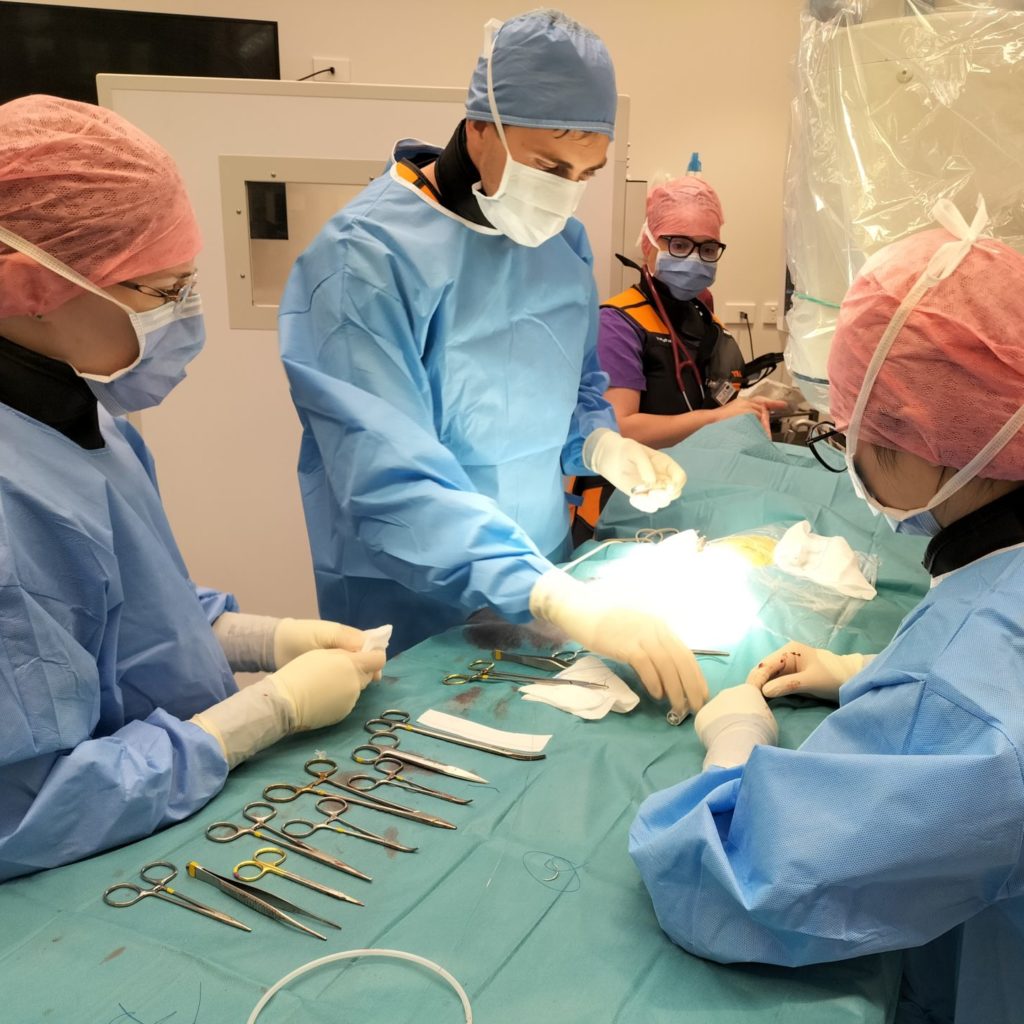
Cardiology at VSA
Specialist Veterinary Cardiology
Watch Dr Keaton Morgan explain more about VSA’s specialist veterinary Cardiology service.
What is VSA Cardiology?
VSA Cardiology is a multi-faceted service centered around the heart; the most vital organ in the body.
We provide advanced cardiology diagnostic services
We provide advanced diagnostic services:
- Echocardiogram:
An Echocardiogram, often referred to as an Echo is an ultrasound of the heart. It gives the cardiologist an image of the inside of the heart to determine how well the heart muscle is pumping, how well the valves are working and whether any defects are present inside the heart.
- CT:
A CT scan is a 3-dimensional x-ray which is available for complex congenital heart disease and surgical planning if required in specific circumstances.
- Electrocardiogram:
An Electrocardiogram is a recording that evaluates the electrical activity of the heart over 5 minutes.
This helps to diagnose the cause of an abnormal heartbeat, called an arrhythmia, and implement the correct treatment.
- 24 Hour Holter Monitor:
Using a holter monitor is a noninvasive method of assessing the heart’s rhythm and rate. It is a 24-hour electrocardiogram that is recorded while a patient is wearing a recorder.
By wearing the monitor for 24 hours it gives us an analysis of the rhythm and rate of the heart throughout the day and night of the patient.
- Loop Recorder:
A loop recorder is a small electrical device that can be implanted under the skin adjacent to the heart. This allows ECG monitoring of the heart rhythm for up to 5 years.
- Angiography:
Angiography uses contrast in the vessels to identify abnormal connections in the vasculature. It is useful to aid the diagnosis and confirm appropriate treatment during minimally invasive procedures.
Some of the most common heart conditions we see in our patients
Patent ductus arterioles (PDA) occlusion
A PDA is a birth defect in the heart caused by incomplete changes in the heart’s circulation when an animal is born. In most cases it is usually picked up in an initial puppy or kitten check-up where a heart murmur is heard.
The ductus arteriosus is an important blood vessel that ensures that blood does not go to the lungs unnecessarily as the fetus is developing in the uterus. During the first few hours after birth, this blood vessel would naturally close off. This allows blood to travel normally through the lungs for oxygenation as the lungs begin to function when the animal takes its first breath. In some baby animals, the ductus arteriosus remains open (patent). This results in serious, life-threatening changes in the way that the heart pumps blood through the heart and to the rest of the body.
Once a patient has been diagnosed with a PDA the goal of treatment is to close the open ductus arteriosus.
At VSA this can be accomplished through cardiac catheter-based (minimally-invasive surgery) occlusion. Catheter based occlusion is a minimally-invasive key-hole procedure and the patients usually go home the following day.
Pulmonic stenosis balloon valvuloplasty
Pulmonic stenosis is a congenital heart defect of the valve that is found between the right ventricle and the pulmonary artery. It is most commonly seen in breeds such as Labradors, Samoyeds, Terriers and brachycephalics.
At VSA we use a technique called balloon valvuloplasty in the treatment for valvular pulmonic stenosis.
This treatment involves the introducing of catheters via the veins and a deflated balloon placed through the abdominal stenotic valve. When the balloon is inflated, it opens the restricted valve leaflets. Measurements of the pressure gradient before and after treatment give information as to the success of the treatment.
Artificial pacemaker placement
An artificial pacemaker is placed in dogs with dangerously slow heart rates because of damage or aging of the conductive tissue.
The artificial pacemaker is able to take control of the heart rate to prevent it getting too slow, or stopping altogether. The pacemaker can also recognise movement to increase the heart rate with exercise. The result is prevention of death from slow/ stopping hearts and improved quality of life.
Here at VSA we have access to the latest pacemaker technology, allowing the pacemakers function and battery life to be monitored remotely.
Cutting balloon procedures
Subaortic stenosis is a narrowing of the area underneath the aortic valve, that causes an obstruction or blockage of the blood flow through the heart. The narrowing can be classified as mild, moderate, or severe. In moderate or severe cases the heart is forced to work harder and potentially be harmful to the heart’s health.
A cutting balloon is an inflatable vascular balloon with micro blades attached. The balloon is carefully positioned across stubborn obstructions through key hole surgery into the peripheral vessels. This type of balloon can be used to open more restrictive obstructions such as severe subaortic stenosis. Subaortic stenosis is a fibrous-muscular narrowing under the aortic valve which in particular cases may respond to a cutting balloon procedure.
Intravascular stenting
Stents are metallic expandable tubes that can be carefully implanted across tough, stubborn obstructions that respond poorly to a balloon inflation. The stent keeps the area open, eliminating the obstruction. This can be used for a range of vascular obstructions within the cardiovascular system, By opening up the region permanently, bloodflow can move freely and not back-up around the body.
Intravascular coiling
Coils are small metallic structures covered in a fine feather -like material. The feathers promote clot formation. Therefore, coils can be used to block abnormal vascular connections and can be delivered in a minimally -invasive procedure through a vessel in the neck or leg.
The most common procedure that requires the use of vascular coils in intra-hepatic portosystemic shunts, however at VSA they can also be used to occlude other abnormal vessels if deemed necessary.
FAQs
My pet has a heart murmur:
A heart murmur is a harsh heart sound caused by turbulent blood flow. Murmurs can be caused by a leaky valve, shunt, or fast blood flow in the heart. To find out the cause of a murmur we perform an ultrasound of heart (also known as an echocardiogram). This test will tell whether the murmur can be ignored, monitored or would benefit from medical or surgical treatment. It is a painless test where an ultrasound probe is placed on the side of the chest while the animal is laying on its side on a table. You can be present during the ultrasound and see the images in real time.
My animal has an irregular rhythm:
An irregular heart rhythm is also called an arrhythmia. This is investigated with an electrocardiogram. This is different than an ECHOcardiogram (which is an ultrasound of the heart), instead the ELECTROcardiogram (or ECG) assesses the electrical activity of the heart. There can be many reasons for a heart rhythm to be irregular, from normal fluctuations to insidious arrhythmias which can result in sudden cardiac death. The ECG is the best first step to assess the rhythm but often you also need to check the structure of the heart with an ultrasound of the heart. We also have the ability to monitor the rhythm for 24 hours while at home (Holter monitor) or if needed, implant a device to monitor the heart rhythm for up to 5 years (Loop recorded).
My puppy/kitten has a murmur:
There are only two general reasons for a puppy or kitten to have a murmur: Physiological (“normal”) or congenital heart disease. Most soft murmurs are physiological which should disappear by 6 months of age. If a soft murmur is heard in your puppy or kitten, it is reasonable to wait and have another auscultation at 6 months. If it is still present at 6 months and it is a moderate/loud murmur or you just want to have the peace of mind and have it checked, in order to diagnose the cause of the murmur you need to perform an echocardiogram (ultrasound of the heart). This is performed in the consult room with you and your pet so you can see the cause of the murmur.
I have a Doberman and have heard they get heart disease:
Unfortunately, Dobermans do have a high prevalence of heart disease with over 50% of dogs developing DCM within their lifespan according to a European study. DCM (Dilated Cardiomyopathy) is a cardiac disease where the heart develops poor contractility and dilates in size. This can lead to consequences such as collapse, heart failure and sudden death. Because of the high prevalence of disease, the recommendation is to have an annual echocardiogram and Holter (24-hour electrocardiogram at home) from the age of 3 years. There are medications that can be given once the first signs of structural heart changes are observed to prolong the onset of heart failure and reduce the risk of sudden cardiac death.